This article is taken from the monthly magazine Sciences et Avenir n°948, dated February 2026.
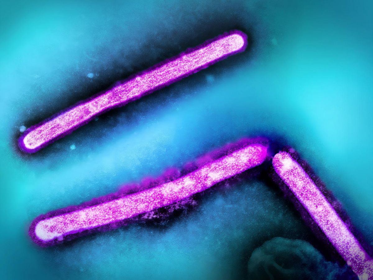
Revealing in real time the infiltration of the flu virus into our cells is what the combination of two powerful imaging tools, atomic force microscopy (AFM) and fluorescence microscopy, has made possible.
Examine the action of antivirals at the cellular level
The unprecedented resolution of this supermicroscopy made it possible to observe how respiratory cells defend themselves before becoming infected. This allows for the examination, at the cellular level, of the action of prescribed antiviral drugs.
These results were obtained by Hokkaido University (Japan) and published in the journal Proceedings of the National Academy of Sciences.
Find the latest articles from Science and Future Regarding imaging techniques:
The Infinitely Small: Techniques for Observing the Invisible
VIDEO. Neuromuscular diseases under imaging
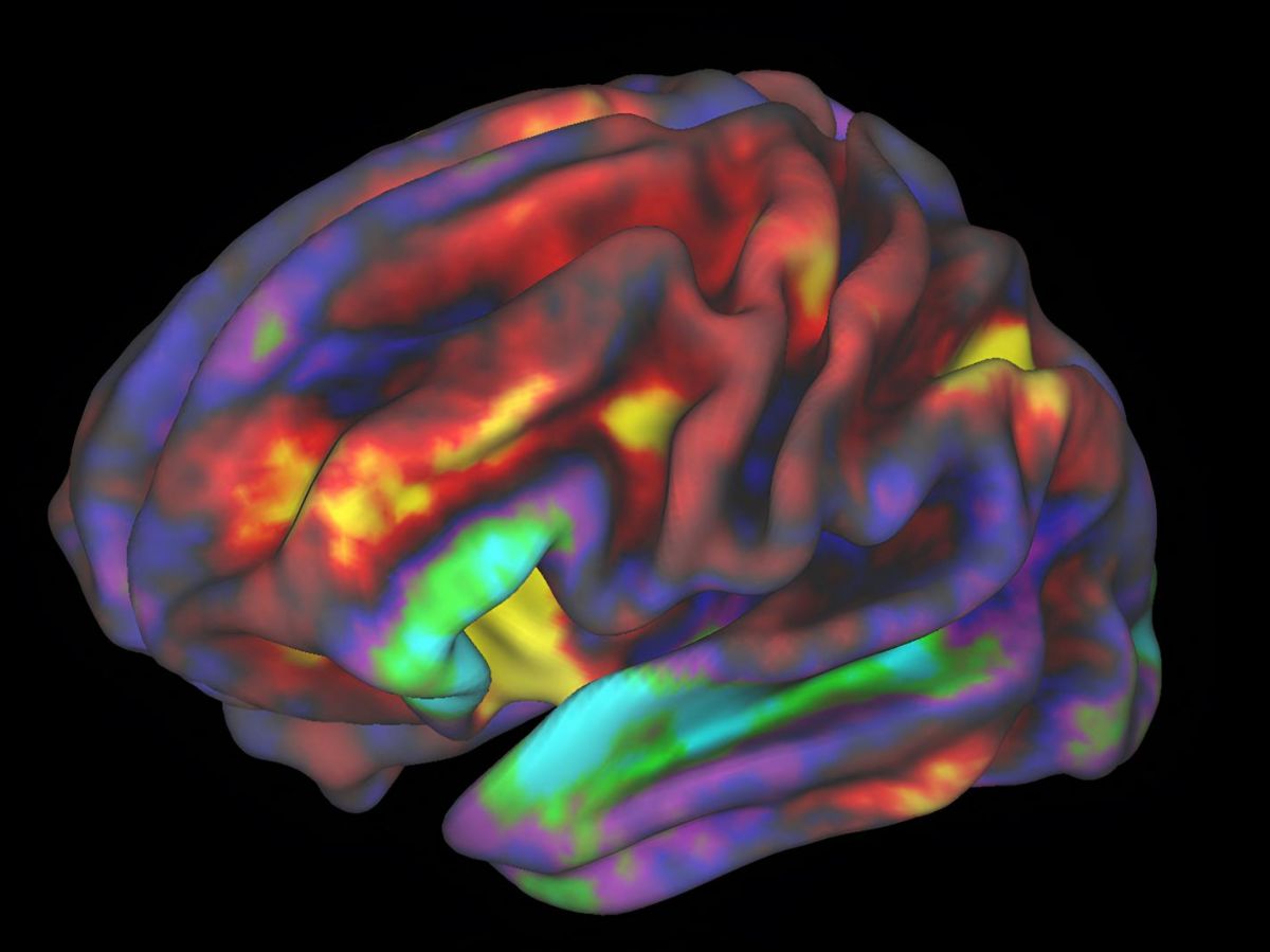
Child's Brain: Images of Unprecedented Precision Obtained by French Researchers