Seeing the blood circulation of an organ in 4D (that is, in a relief that evolves in real time) is now possible. Proof of this is the first-ever ultra-precise images of the microcirculation of an entire organ, just published in the review Nature.
An observation that ranges from large vessels to "precapillary arterioles"
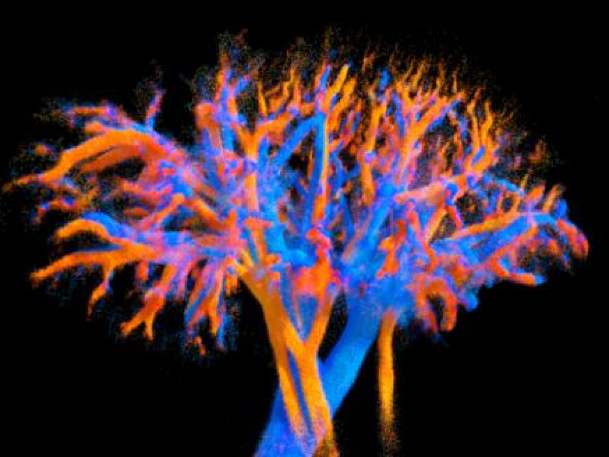
This pioneering work, carried out to date only in animals, is the result of the work of a team of researchers from the Institute of Physics for Medicine (Inserm/ESPCI Paris-PSL/CNRS). Scientists used a new ultrasonic probe combining several lenses to obtain a wide field of view. It was developed as part of Nabil Haidour's doctoral research under the supervision of Clément Papadacci. This device enabled to dynamically map the complex network of blood vessels that carry blood to tissues and organs. And theThe result is quite stunning, with images of vascular tree formations emerging live in front of the viewer.

This work represents a first in the field of imaging, as until now no method allowed for the visualization of microcirculation in 4D and at the scale of an entire organ. Here, pig hearts, kidneys, and livers were mapped with unprecedented precision.
The objective is obviously not simply to see, but above all to analyze and evaluate the integrity of the circulatory system as a whole, from the large arteries (the largest being the aorta with a diameter of 2.5 cm) down to the smallest arterioles (less than 100 micrometers). “ Our approach allows for a more in-depth analysis of hemodynamics, from large vessels to precapillary arterioles, by providing large and rich datasets on whole-organ vascularization., write the authors in their abstract.
Facilitating the diagnosis of certain pathologies related to blood circulation

“ It could also contribute to advancing the diagnosis of microcirculatory disorders and the monitoring of treatments for small vessel diseases, diseases for which diagnosis is complex and is made by excluding other pathologies., explains the first author, Clément Papadacci. Because that is precisely the point of this new imaging technique which must now move to the clinical stage, with a trial planned involving around forty people.
This development could both allow for a better understanding of the circulatory system (veins, arteries, vessels and lymphatic system), and also facilitate the diagnosis of certain pathologies related to blood circulation. This work will be carried out with the help of ART Biomedical Ultrasound, a technological research accelerator created by Inserm and integrated into the Institute of Physics for Medicine. "The probe can be connected to a small, portable device, allowing its integration into medical practice."Clément Papadacci predicts this.