Until now, only snapshots of this crucial moment existed. But for the first time, the implantation of a human embryo has been filmed in real time and in 3D by a team of researchers. Beyond witnessing a unique moment in conception, these images also provide insight into how embryos behave. They reveal that they are far less passive at the heart of this process than previously thought.
After fertilization, when the egg and sperm meet, the embryo begins to divide and travel toward the uterus. About 5 to 7 days after fertilization, it arrives in the uterus and attaches to the uterine lining, also called the endometrium. This key step for the continuation of the pregnancy is called implantation. It allows the embryo to position itself as close as possible to the mother's blood vessels, to which the placenta will later connect.
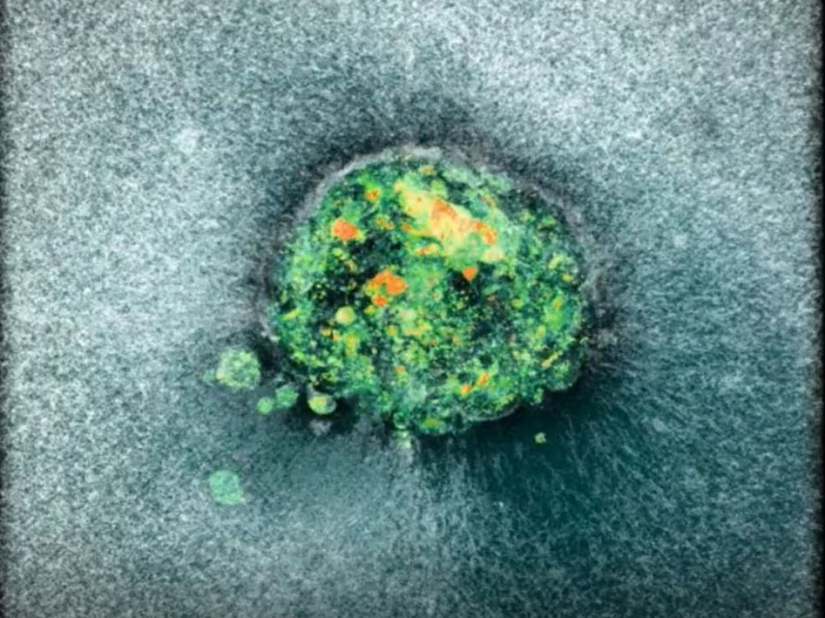
This short clip shows images of the embryo, its contours shifting as it moves through the uterine tissue. Collagen fibers can also be seen being pulled along by the embryo as it passes through. Video credit: Sarah Moreira Castro
In the image, we see the embryo exerting a force on the uterus, a real " effort " in order to implant itself. And behind it, the trace of its passage: the uterine fibers that it eliminated to continue its progression. " We have noticed that human embryos literally burrow into the uterus, exerting considerable force in the process. This movement is necessary for the embryo to enter and integrate into the uterine tissue.", tells Science and Future Amélie Godeau, researcher at the Institute for Bioengineering of Catalonia (IBEC) in Spain and first author of the study published in Science Advances. “ Perhaps the contractions or, let's say, tensions felt by some women at the time of embryo implantation could come from this movement, when the embryo moves in the womb and reorganizes it.. »

Once the implantation site is chosen, the embryo releases enzymes designed to destroy the surrounding tissues, making it easier for it to pass through. Here again, it also exerts a force to penetrate deeper into the uterus. This is because these fibrous tissues are made of collagen, a rigid protein that our tendons and cartilage are also made of. The embryo must degrade the collagen, we can clearly see where it has passed and where the enzymes have digested these collagen molecules. » The rest of the collagen fibers move along its path, until the embryo reaches its goal: it penetrates deep into the tissues to the maternal blood vessels.
An artificial uterus made of collagen
To achieve this feat, the team developed an artificial uterus made of collagen fibers. It is the most common extracellular protein in humans. The artificial uterus must achieve the right pH and ensure that the embryos survive.", explains Amélie Godeau. Everything is done to recreate the natural environment of an embryo. " The human embryos used for these experiments were obtained under very strict conditions. These are embryos from couples who have completed their in vitro fertilization (IVF) process and who are free to donate these embryos to science, donate them, or keep them. The embryos we received were donated to science.", explains the researcher. In Spain as in France, research on embryos is limited to 14 days of life for ethical reasons.
This work was launched precisely to better understand how reproduction works. Problems with embryo implantation remain one of the main causes of infertility. One-third of embryos never implant, and one-third detach immediately after implantation, making the pregnancy undetectable. In total, these are two-thirds of pregnancies for which implantation is a limiting factor.", recalls Amélie Godeau. By better observing how this process takes place, the team hopes to advance research into infertility. In the meantime, they will have already allowed us to witness – for the first time – one of the crucial moments in reproduction.