You are here
Press release
Thursday, February 27, 2025

NIH study reveals key players underlying disease onset and repair
Using an animal model of multiple sclerosis (MS), researchers at the National Institutes of Health (NIH) have created a four-dimensional brain map that reveals how lesions similar to those seen in human MS form. These results, published in Science, open a window into the early state of the disease and could help identify potential targets for MS treatments and brain tissue repair.
The researchers, led by postdoctoral researcher Jing-Ping Lin, Ph.D., and principal investigator Daniel S. Reich, MD, Ph.D., both at the NIH's National Institute of Neurological Disorders and Stroke (NINDS), combined repeated MRI imaging with brain tissue analysis, including gene expression, to track the onset and development of MS-like lesions. They discovered a new MRI signature that can help detect brain regions at risk for damage weeks before visible lesions appear. They also identified "microenvironments" within the affected brain tissue based on observed patterns of neuronal function, inflammation, immune and support cell responses, gene expression, and levels of damage and repair.
“Identifying the earliest events that occur after inflammation and distinguishing between those that are reparative and those that are damaging can potentially help us identify MS disease activity earlier and develop treatments to slow or stop its progression,” said Dr. Reich.
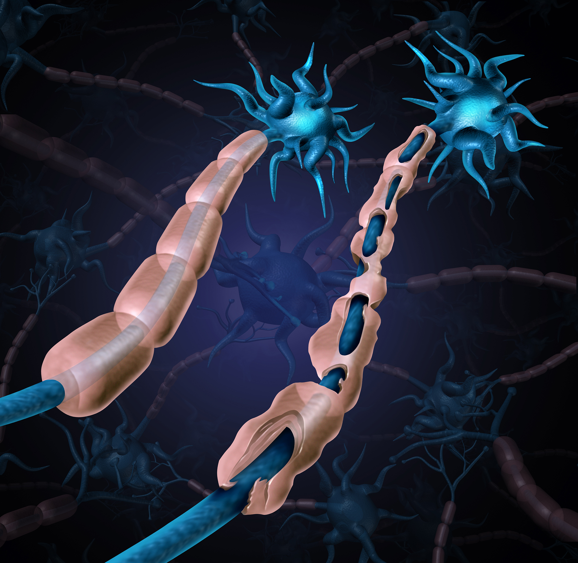
MS is caused by the body's immune system attacking the protective sheath of nerve fibers, called myelin. This leads to inflammation, loss of myelin, and the formation of "lesions" or "plaques" in brain tissue. Most of what we know about MS progression comes from analyzing postmortem human brain tissue, usually obtained decades after the initial onset of the disease. This means that early changes that occurred before symptoms appeared are missed.
To mimic conditions in the human brain, the researchers chose not to use a mouse model for MS, but instead to advance a model that uses the marmoset, a non-human primate. Compared to the mouse brain, both the marmoset and the human brain have a higher ratio of white matter (the brain's "wires") to gray matter (the neuronal cell bodies). The marmoset model creates multiple lesions that closely resemble those seen in human MS and can be tracked in real time using MRI imaging. Because these lesions can be induced experimentally, the model offers insight into the early stages of inflammation and immune responses that lead to MS-like demyelination.
One of the key players identified was a specific type of astrocyte, one of the brain's supporting cell types, which activates a gene called SERPINE1 or plasminogen activator inhibitor-1 (PAI1). They found SERPINE1-expressing astrocytes in the vulnerable edges of the brain before visible damage occurs, clustering near blood vessels and fluid-filled ventricles of the brain and signaling future areas of injury development. These astrocytes also appear to influence the behavior of other cells near the area of injury, including the ability of immune cells to enter the brain and contribute to inflammation, as well as precursor cells involved in myelin repair.
Given that SERPINE1-expressing astrocytes accumulate at the edges of growing lesions, where damage occurs but also healing begins, their potential dual role in coordinating signals that could lead to either tissue repair or further damage was an unexpected wrinkle that will require further study. It is possible that the early responses are part of a protective mechanism that becomes overwhelmed as the injury progresses. It is also possible that the same mechanism could itself become disease-causing.
"If you imagine a fort under siege, the walls might initially hold back the attack," Dr. Reich said. "But if those walls are breached, all the defenses inside can be turned against the fort itself."
These findings may also have implications for brain injury beyond what is seen in MS. Although there are different types of focal brain injury, including traumatic brain injury, stroke, inflammation, and infection, there are a limited number of ways in which tissues can respond to injury. In fact, many of the responses observed here to inflammation, stress, and tissue injury are likely common to all types of injury, and the brain map created in this study can serve as a resource to enable comparisons in a more human context.
The scientific teams are building a new model of another autoimmune disease affecting the brain's boundaries. They are also looking to expand their dataset to include older animals, which could help improve our understanding of progressive MS, a pathological condition with a significant unmet therapeutic need.
This study was funded in part by the NIH Intramural Research Program with additional support from the Dr. Miriam and Sheldon G. Adelson Medical Research Foundation and the National Multiple Sclerosis Society.
About the National Institute of Neurological Disorders and Stroke (NINDS): The NINDS is the nation's largest funder of brain and nervous system research. NINDS's mission is to seek fundamental knowledge about the brain and nervous system and to use this knowledge to reduce the burden of neurological disease.
About the National Institutes of Health (NIH): NIH, the nation's medical research agency, comprises 27 institutes and centers and is part of the U.S. Department of Health and Human Services. NIH is the primary federal agency that conducts and supports basic, clinical, and translational medical research, studying the causes, treatments, and cures for common and rare diseases. For more information about NIH and its programs, visit www.nih.gov.
NIH…Transforming Discovery into Health®
Article
Lin JP et al. 4D brain map of the marmoset reveals MRI and molecular signatures for the occurrence of multiple sclerosis-like lesions. Science. February 28, 2025. DOI: 10.1126/science.adp6325
###