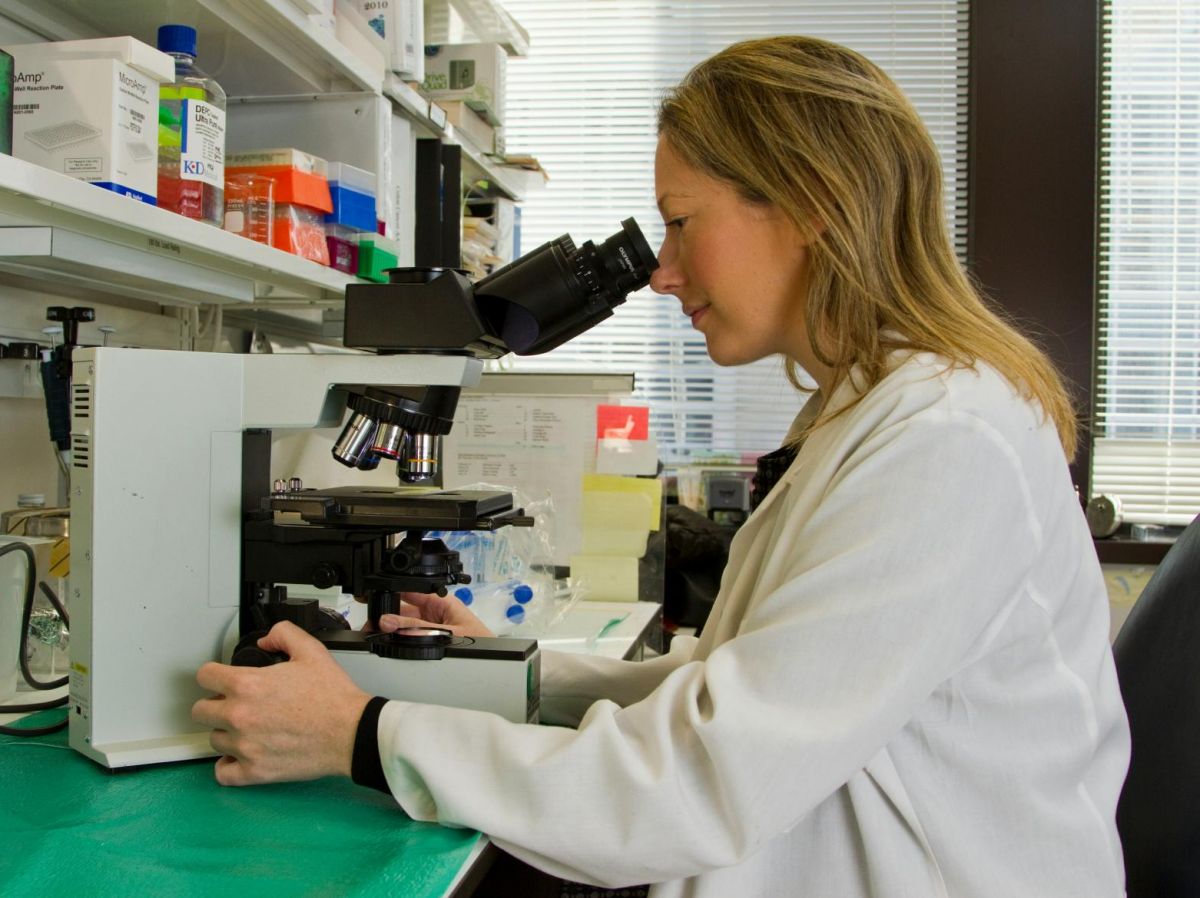
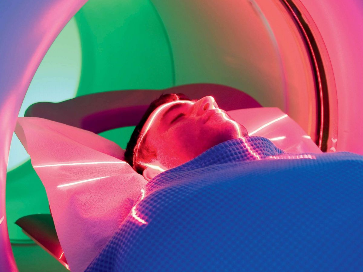
"I have noticed, both through my personal relationships and in the context of my work, that Many people avoid getting diagnosed for fear of the pain of biopsies., points out Ciro Chiappini, lecturer in nanomaterials and biointerfaces at King's College London (United Kingdom), during an interview with Science and FutureA major problem for patients whose condition can worsen without early detection.
Based on this observation, he and his team propose a painless method to obtain the same type of information as a biopsy, without fear. Their prototype of a painless patch made of tens of millions of nano-needles is presented in the journal Nature Nanotechnology.
Needles 1,000 times thinner than a hair
A biopsy involves removing a sample of tissue or organ, often using a special needle, scalpel, brush, or other suitable instrument. The sample may be taken under imaging guidance for greater precision. It is then analyzed, and the results are useful for various types of diagnoses, particularly cancer.
While every effort is made to ensure that the examination, although invasive, is as painless as possible, it is common to be apprehensive, or even avoid it. Alternatives exist, such as liquid biopsies, which use blood or saliva instead of taking a tissue sample. This is a much less invasive technique and can provide a wealth of information. But in most cases, it still requires a blood sample, a procedure that is also dreaded.
The patch format offers a painless and less invasive alternative. The nanoneedles are 1,000 times thinner than a human hair and do not remove tissue, making the process less intimidating for patients than traditional biopsies.
An analysis without tissue sampling
In preclinical studies, the team applied the patch to brain cancer tissue taken from human and mouse biopsies. The nanoneedles extracted molecular fingerprints—including lipids, proteins, and mRNA—from the cells without damaging the tissue.
This fingerprint is then analyzed using mass spectrometry (a technique that measures the mass of molecules to identify them) and artificial intelligence, providing healthcare teams with detailed information on the possible presence of a tumor, its response to treatment and the progression of the disease at the cellular level. The patches do not remove any tissue and allow multiple tests to be performed on the same area, something that was previously impossible with conventional biopsies.
The only challenge remains reaching certain organs. If we're trying to reach deep tissues, we need to find a way to access them. Researchers are exploring ways to use existing procedures and add nanoneedles, such as endoscopy (a flexible tube equipped with a camera to observe the digestive system), for example. This way, no additional surgery would be necessary.
The patches could also be used during brain surgery, to guide real-time decisions about removing cancerous tissue. “By applying the patch to a suspect area, analyses would be obtained in 20 minutes.”, says the lead author. The patch is removed immediately after the exam, but the needles are designed to degrade completely within a few days if any residue remains.
"This could mark the beginning of the end of painful biopsies."
"Patients would be more likely to consult a doctor if they were not afraid of a biopsyThis could mark the beginning of the end of painful biopsies and late detection., adds the researcher.
Nanoneedles are already widely used in healthcare. "We currently use them to administer things like mRNA and CRISPR for gene editing, and they are very promising”. Intraocular lenses with nanoneedles are used to detect certain eye pathologies, such as Fuchs' dystrophy. For skin, there are nanoneedle plasters.
Many of these projects are in clinical trials. If all goes well, nanoneedle patch biopsies could be used clinically within the next few years.


