Scientists map unprecedented details of connections and visual perception in the mouse brain
April 9, 2025
Media Advisory
Wednesday, April 9, 2025

An NIH-funded project is helping to untangle the brain's wiring, providing clues about how we see the world.

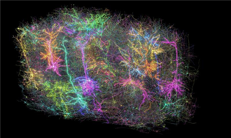
This image shows a subset of over 1,000 of the 120,000 brain cells reconstructed in the MICRONS project. Each reconstructed neuron is a different random color. This is a symbolic representation of the entire dataset. There are many more recorded neurons than glowing neurons, and many more reconstructed neurons than were included in the rendering. Allen Institute
What

As part of a massive scientific effort Funded by the National Institutes of Health (NIH), hundreds of researchers helped map the connections between hundreds of thousands of neurons in the mouse brain, then overlaid their firing patterns in response to visual stimuli. This fundamental scientific advance is essential for understanding how our brains process visual information to reconstruct the images we see every day.
Information processing in the human brain occurs through the electrical activation of 86 billion neurons that make billions of connections between them. The secrets of how our brains allow us to think, feel, and act lie in the complexity of their wiring and the stream of electrical signals that pass through them in milliseconds. Although the current findings focus on a tiny part of the brain, they reveal the intricate connections between cells and show how these connections are wired to produce functional responses. This previously inaccessible information could help us understand normal brain function and identify dysfunctions resulting from various disorders or injuries.
To conduct this study, the researchers presented video clips to mice genetically engineered to make their neurons emit light when they fire. The firing patterns of neurons in areas of the brain's surface associated with vision were optically recorded on a cubic millimeter, about the size of a grain of sand. At the heart of this surprisingly small amount of tissue lies a remarkable complexity: four kilometers of axons, the processes that nerve cells use to communicate with each other, intertwine to form more than 524 million connections called synapses across more than 200,000 cells.
To map these connections, the teams worked 12 hours a day for 12 consecutive days to carefully cut and image ultrathin slices of brain tissue using electron microscopes (EM). Reconstruction was the next most challenging step, requiring the precise stitching of nearly 28,000 EM images to align the connections spanning the volume of brain tissue. This was followed by months of tracing the connections using deep learning algorithms, followed by manual and automated review. Predictive deep learning models explaining visual information processing in the cortex were built and validated. In total, the amount of data collected to create this tiny map amounts to 1.6 petabytes—roughly the equivalent of 22 years of continuous HD video.
These findings come at a time when maps of neurons and their connections are increasingly revealing the mysteries of the brain. In 2023, research funded by the "Brain Research Through Advancing Innovative Neurotechnologies" initiative ®” from the National Institutes of Healthor "NIH BRAIN Initiative®", produced the first complete cell atlas of the mouse brain, including the types and locations studied in more than 32 million cells. Last year, the NIH BRAIN Initiative's "Flywire" project led to the complete mapping of the brain of the common fruit fly, demonstrating the unique value of mapping the entire brain.
Funding for this project was provided by the Intelligence Advanced Research Projects Activity's Machine Intelligence from Cortical Networks (MICrONS) program and the NIH BRAIN initiative. The results, published in a set of 10 articles in the family of journals Nature represent more than seven years of work carried out by more than 150 scientists from around the world.
The mouse connectome data detailed in this press release can be viewed online using the resource MICRONS Explorer.
Who
John Ngai, Ph.D., director of the NIH BRAIN Initiative, is available for comment.
Article
The articles from the MICrONS et al. consortium are available here.
The NIH BRAIN Initiative, a multidisciplinary collaboration bringing together 10 NIH institutes and centers, is uniquely positioned to make cross-disciplinary discoveries in neuroscience to revolutionize our understanding of the human brain. By accelerating the development and application of innovative neurotechnologies, the BRAIN Initiative® enables researchers to understand the brain at unprecedented levels of detail in both health and disease, thereby improving how we treat, prevent, and cure brain disorders. The BRAIN Initiative involves a multidisciplinary network of federal and non-federal partners whose current missions and research portfolios complement the goals of the NIH BRAIN Initiative.
About the National Institute of Neurological Disorders and Stroke (NINDS): The NINDS is the nation's leading funder of brain and nervous system research. Its mission is to develop fundamental knowledge about the brain and nervous system and use it to reduce the burden of neurological disease.
About the National Institutes of Health (NIH): The NIH, the nation's medical research agency, comprises 27 institutes and centers and is part of the U.S. Department of Health and Human Services. The NIH is the primary federal agency conducting and supporting basic, clinical, and translational medical research, and investigating the causes, treatments, and cures for common and rare diseases. For more information about the NIH and its programs, visit www.nih.gov.
NIH…Transforming Discovery into Health®
###