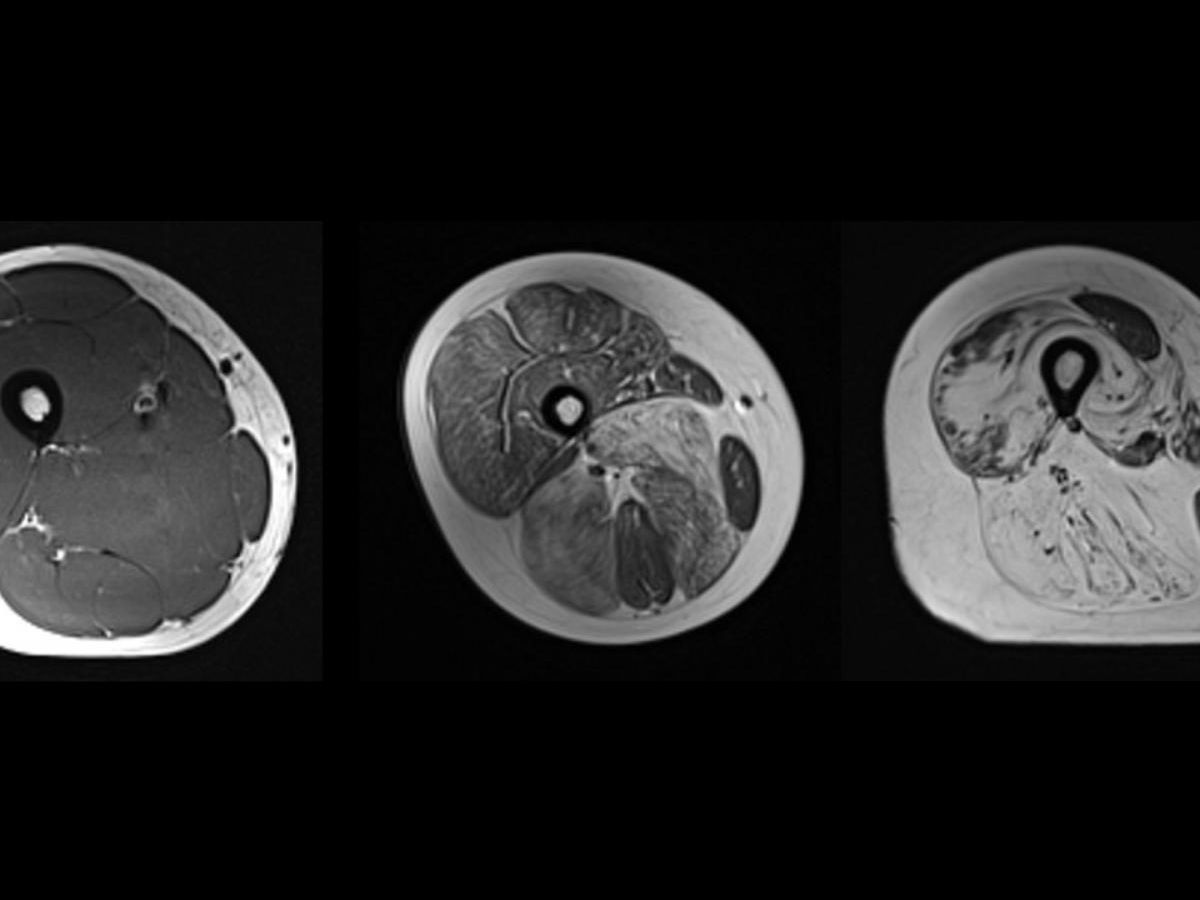

At the Institute of Myology, a muscle expertise center created by the AFM – Téléthon located in the heart of the Pitié Salpêtrière Hospital, the nuclear magnetic resonance (NMR) imaging and spectroscopy laboratory studies cases of neuromuscular diseases and develops innovative muscle imaging methods. They allow several factors to be precisely quantified and thus the development of the disease to be better monitored, depending on whether it is treated or not, such as the occurrence of intramuscular fat which ultimately prevents the muscles from functioning, necrosis or edema.
An interview with Benjamin Marty, co-director of the imaging laboratory and NMR spectroscopy
Among the most well-known and studied diseases: Duchenne muscular dystrophy, a genetic disease in children that causes progressive weakening to the point of affecting the cardiac and respiratory muscles. Research is particularly active on ways to study these muscles, which are extremely small and impossible to immobilize during an examination.
Find the interview with Benjamin Marty, co-director of the imaging laboratory and of NMR spectroscopy, which explains Science and Future How to visualize neuromuscular diseases under imaging.
