NIH researchers improve common clinical device to better see the back of the eye
April 23, 2025
Press release
Wednesday, April 23, 2025
A new technique allows the retina to focus more sharply.

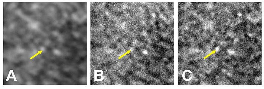
Comparison of the same retinal area labeled with indocyanine green and viewed in 3 different ways. A) Scanning laser ophthalmoscopy. B) AI-enhanced scanning laser ophthalmoscopy. C) Adaptive optics scanning laser ophthalmoscopy. Arrows highlight the same cell viewed in different modalities.
Scientists at the National Institutes of Health (NIH) have harnessed artificial intelligence to transform a device designed to view tissues at the back of the eye into one sharp enough to distinguish individual cells. This technique offers imaging resolution that rivals the most advanced devices available and is cheaper, faster, and does not require specialized equipment or expertise. This strategy has implications for early disease detection and for monitoring treatment response by making visible what was once invisible.
“AI potentially puts next-generation imaging into the hands of standard eye clinics. It’s like adding a high-resolution lens to a basic camera,” said Johnny Tam, Ph.D., a researcher at the NIH’s National Eye Institute and lead author of the study, published in Communications Medicine.
Imaging devices called ophthalmoscopes are widely used to examine the light-sensitive retina at the back of the eye. A scanning laser ophthalmoscope is standard in eye clinics, but its resolution can only distinguish tissue-level structures, such as lesions, blood vessels, and the optic nerve head. Newer-generation ophthalmoscopes with adaptive optics (a technology that compensates for light distortion) can distinguish cellular features, providing better diagnostic information. However, adaptive optics imaging is still in the experimental phase.
Tam and his colleagues developed a custom AI system to digitally enhance images of a layer of tissue located beneath the light-sensitive photoreceptors, called the retinal pigmented epithelium (RPE). The first step was to teach the system to distinguish between poor, average, and good image quality. To do this, the researchers provided the system with more than 1,400 images of different areas of the retina, obtained using adaptive optics ophthalmoscopy. They then provided the system with corresponding images from the same retinal areas, but obtained using standard ophthalmoscopy. A test of image sharpness showed that the AI increased clarity eightfold.
“Our system used lessons learned from evaluating adaptive optics images to digitally enhance images obtained by standard ophthalmoscopy,” Tam said. “It’s important to emphasize that the system isn’t creating something out of thin air. The features we see in RPE cells with standard imaging are there, but they’re blurry.”
These techniques involve injecting a dye called indocyanine green (ICG) into the bloodstream to increase the contrast of anatomical features. In ophthalmic clinics, ICG is typically used to image the blood vessels of the eye.
“Our ICG imaging strategy enables rapid and systematic assessment of retinal pigment epithelium (RPE) cells in the clinic,” said Joanne Li, Ph.D., first author of the report and a biomedical engineer in Tam’s lab. “Using AI, high-quality images of RPE cells can be obtained in seconds, using standard clinical imaging instruments.”
The function of RPE cells is to nourish and support photoreceptors. Various blinding pathologies primarily affect RPE cells, including age-related macular degeneration, vitelliform macular dystrophy, and Stargardt disease. However, RPE cells cannot be easily imaged clinically. AI-enhanced ICG ophthalmoscopy brings RPE imaging within reach of a conventional eye clinic.##
This press release describes a basic research finding. Basic research improves our understanding of human behavior and biology, which is essential for developing new, more effective methods for preventing, diagnosing, and treating disease. Science is an unpredictable and incremental process: every scientific advance builds on previous discoveries, often in unexpected ways. Most clinical advances would not be possible without the knowledge gained from basic research. To learn more about basic research, visit https://www.nih.gov/news-events/basic-research-digital-media-kit.
The NEI leads the federal government's efforts to eliminate vision loss and improve quality of life through vision research… by stimulating innovation, fostering collaboration, expanding the vision workforce, and educating the public and key stakeholders. The NEI supports basic science and clinical programs aimed at developing sight-preserving treatments and expanding opportunities for people with low vision. For more information, visit https://www.nei.nih.gov.
About the National Institutes of Health (NIH): The NIH, the nation's medical research agency, comprises 27 institutes and centers and is part of the U.S. Department of Health and Human Services. The NIH is the primary federal agency conducting and supporting basic, clinical, and translational medical research, and investigating the causes, treatments, and cures for common and rare diseases. For more information about the NIH and its programs, visit www.nih.gov.
NIH…Transforming Discovery into Health®
References
Li J, Liu J, Das V, Le H, Aguilera N, Bower Aj, Giannini JP, Lu R, Abouassali S, Chew EY, Brooks BP, Zein WM, Huryn LA, Volkov A, Liu T, Tam J “Artificial intelligence-assisted clinical fluorescence imaging achieves in vivo cellular resolution comparable to that of adaptive optics ophthalmology." Published on April 28, 2025, Communications Medicine
###